Slopeiomyces

Slopeiomyces cylindrosporus (D. Hornby, Slope, Gutter. & Sivan.) Klaubauf, Lebrun & Crous, Stud. Mycol. 79: 103. 2014. (Type species).

Ascomata perithecial, superficial, globose, black, solitary to gregarious, 290–565 μm diam with a cylindrical, black neck, up to 242 μm long, 48–116 μm wide at base. Ascomatal wall up to 54 μm thick. Paraphyses unbranched, septate, hyaline. Asci 8-spored, unitunicate, clavate, straight to curved, 65–135 × 9–16 μm, with a refractive apical ring. Ascospores cylindrical, fusoid, to sigmoid, 3–8-septate, slightly curved, hyaline, 37–69 × 3.2–5.6 μm. Asexual state phialophora-like. Conidiogenous cells phialidic, subcylindrical to ampulliform, straight or curved, 6.3–19.6 × 2.4–4.2 μm, hyaline, with a flared collarette. Conidia ellipsoidal, fusiform, cylindrical to lunate, straight to curved, aseptate, hyaline, 2.9–6.3 × 0.7–2 μm (Description from Hornby et al., 1977).

Typification: Holotype IMI192606. Ex-holotype cultures CBS609.75, CBS610.75, CBS611.75.

Gene sequences: NG061048 (18S), JX134667 (ITS), NG057751 (28S), JX134707 (MCM7), JX134721 (RPB1), JX134693 (TEF1).

Genome sequences: SRX798639 (transcriptome).

Hosts/substrates: From roots of Poaceae.

Distribution: UK, USA.

Pseudotracylla

Pseudotracylla dentata B. Sutton & Hodges, Nova Hedwigia 27(3-4): 699. 1976. (Type species).

Conidiomata pycnothyrial, superficial, solitary to gregarious, circular, dark brown, composed of upper and lower walls connected by a central columella; upper wall composed of one layer of conidiogenous cells; lower wall membranous, dark brown. Conidiogenous cells phialidic, smooth, 4.5–8.5 × 3–4 μm, restricted to upper wall and facing down towards lower wall, with a minute collarette. Conidia fusiform to falcate, aseptate, hyaline, smooth, 9.5–12 × 3–4 μm. Sexual state unknown (Description from Sutton and Hodges Jr, 1976).

Typification: Holotype IMI196483K.

Gene sequences: Unknown.

Hosts/substrates: On leaves of Eucalyptus citriodora (Myrtaceae).

Distribution: Brazil.

Pseudotracylla falcata Carris, Mycologia 84(4): 534. 1992.

Conidiomata pycnothyrial, superficial, solitary to gregarious, circular, dark brown, 50–100 µm, composed of upper and lower walls connected by a central columella; upper wall composed of one layer of conidiogenous cells; lower wall membranous, dark brown; columella cylindrical, aseptate, dark brown, 22–26.4 × 4.4–8.8 μm. Conidiogenous cells phialidic, subglobose to cylindrical, yellowish to brown, smooth, 4–10 × 3.5–8 μm. Conidia falcate, aseptate, hyaline, smooth, 16–18 × 1.8–2.6 μm. Sexual state unknown (Description from Carris, 1992).

Typification: Holotype WSP69554. Isotype IMI352168.

Gene sequences: Unknown.

Hosts/substrates: On dead leaves and rotting fruit of Vaccinium macrocarpon (Ericaceae).

Distribution: USA.

Pyriculariopsis

Pyriculariopsis parasitica (Sacc. & Berl.) M.B. Ellis, Dematiaceous Hyphomycetes: 207. 1971. (Type species).
MycoBank: MB 322173.
Helminthosporium parasiticum Sacc. & Berl., Revue mycologique, Toulouse 11(no. 44): 204. 1889.
= Pyricularia musae S. Hughes, Canadian Journal of Botany 36 (6): 800. 1958.

On CMA with sterile rice seeds, hyphae branched, septate, hyaline to brown, smooth, 2–4.5 μm diam. Conidiophores solitary, erect, unbranched, subcylindrical, straight or curved, medium brown, thick-walled, smooth, 5–10-septate, 95–200 × 8–13.5 μm. Conidiogenous cells integrated, terminal, medium brown, smooth, 30–45 × 5–7.5 μm, forming a rachis with several protruding denticles, 2–3.5 × 2–4.5 μm, and a minute marginal frill. Conidia solitary, obclavate, guttulate, 3-septate, smooth, unequally colored, with brown median cells, olivaceous to subhyaline apical and basal cell, 45–65 × 8–10.5 μm; hilum truncate, slightly protruding, with marginal frill, unthickened, not darkened, 2–3 μm diam; apex tapering, subacutely rounded, with a mucoid cap, 3–5 μm diam. Sexual state unknown.

Colonies on PDA 9 cm diam after 7 days at 25 °C in dark; surface flat, transparent with dark mouse-grey areas; reverse with dark spots. Colonies on MEA 5 cm diam after 7 days at 25 °C in dark; aerial mycelium, white, mouse-grey in center, raised, cottony, round; reverse dark mouse-grey in center. Colonies on CMA and OA 9 cm diam after 7 days at 25 °C in dark; surface transparent, with thin, spreading mycelium with scattered dark spots of sporulation (Colony description from Klaubauf et al., 2014).

Typification: Unknown.

Gene sequences: DQ341514 (28S).

Specimens examined: China, Hong Kong, Discovery Bay, Lantao Island, on leaves of Musa, 5 Oct. 1999, K.D. Hyde, HKUCC5562 (Maew HK1, CBS114793).

Hosts/substrates: On leaves of Musa (Musaceae).

Distribution: China.

Copyright 2022 by The American Phytopathological Society. Reproduced, by permission, from Luo, J., and Zhang, N. 2022. The Rice Blast Fungus and Allied Species: A Monograph of the Fungal Order Magnaporthales (https://my.apsnet.org/APSStore/Product-Detail.aspx?WebsiteKey=2661527A-8D44-496C-A730-8CFEB6239BE7&iProductCode=46826). American Phytopathological Society, St. Paul, MN.


Omnidemptus

Omnidemptus affinis P.F. Cannon & Alcorn, Mycotaxon 51: 483. 1994. (Type species).

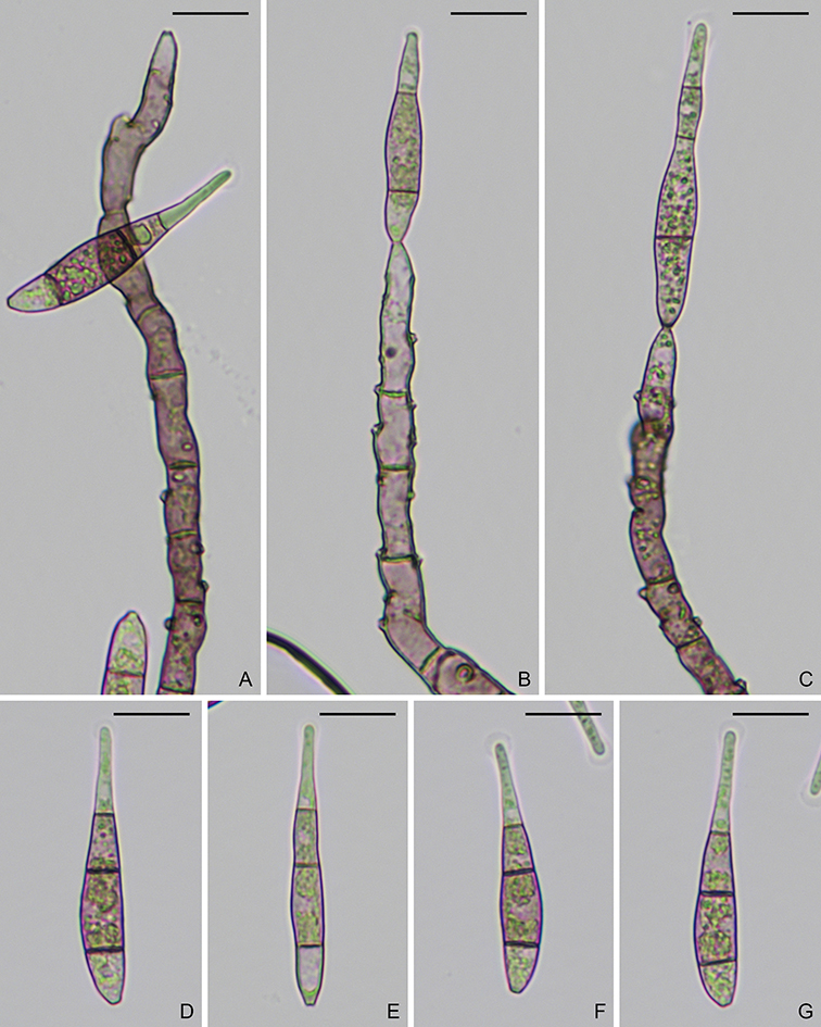
Ascomata perithecial, solitary to gregarious, immersed, globose to subglobose, black, 145–205 μm diam, with a cylindrical, long neck, 100–250 μm long, 80–110 μm at base, 40–60 μm at apex. Paraphyses hyaline, septate, 3–6 μm wide. Asci 8-spored, unitunicate, cylindrical to clavate, 54–68 × 10–13 μm, with an apical ring, 2–3 × 4 μm. Ascospores biseriate, fusiform, curved, 1- or 3-septate, hyaline, smooth, 24.5–33 × 4–5.5 μm. In culture, hyphae septate, hyaline to light brown, smooth, 2–5.5 μm diam. Conidiomata irregularly shaped, dark brown to black, 50–160 μm diam. Conidiogenous cells ampulliform, cylindrical to ellipsoid, dark brown, 11–22 × 6–10 μm, with a cylindrical to flared collarette, 3–4 μm diam. Conidia falcate, 1–2-septate, hyaline, smooth, 21–30 × 3–4 μm. Hyphopodia lobed, olivaceous brown, smooth, 7.5–14 × 5–8.5(–11) μm (Description from Cannon and Alcorn, 1994).

Typification: Holotype BRIP17195. Isotype IMI353435. Ex-holotype culture BRIP17195 (CBS141031, ATCC 200212).

Gene sequences: JX134660 (18S), JX134674 (ITS), JX134686 (28S), JX134714 (MCM7), JX134728 (RPB1), JX134700 (TEF1).

Genome sequences: SRX798629 (transcriptome).

Hosts/substrates: From leaf spots of Panicum effusum (Poaceae).

Distribution: Australia, Spain.

Omnidemptus graminis Hern.-Restr., Gene & Guarro, Persoonia 42: 223. 2019.

Hyphae septate, hyaline to light brown, smooth, 1–3 μm diam. Conidiomata absent or sporodochium-like, irregularly shaped, diffuse, punctiform, dark brown to black. Conidiogenous cells ampulliform to subglobose, brown, smooth, 10–14 μm diam, with a cylindrical collarette, 1 × 3 μm. Conidia falcate, (0–)1-septate, hyaline, smooth, 11–23 × 3–4 μm. Hyphopodia lobed, brown, smooth, 10–15 × 7.5–10 μm, with 1–2 germ pores, 1–3 μm diam. Asexual state unknown.

Colonies on MEA 1.8–2 cm diam after 7 days at 25 °C; surface elevated, cottony, vinaceous buff; margin uneven; reverse black in center, white at periphery. Colonies on OA 3–4 cm diam after 7 days at 25 °C; surface zonate, center cottony, pale mouse grey, periphery glabrous, mouse grey; margin diffuse; reverse mouse grey (Description from Hernandez-Restrepo et al., 2019).

Typification: Holotype CBSH-21887. Ex-holotype culture CBS138107 (FMR12415).

Gene sequences: MK487758 (ITS), MK487734 (28S), MK495980 (TEF1).

Hosts/substrates: On leaves of grasses.

Distribution: Spain.

Omnidemptus lunatus (B. Sutton & Alcorn) Hern.-Restr., J.D.P. Bezerra & Crous, Persoonia 42: 223. 2019.

Conidiomata sporodochium, irregularly shaped, diffuse, light to medium brown. Conidiogenous cells ampulliform, doliiform to lageniform, light brown, smooth, 10.5 –13 × 6.5–11 μm, with a cylindrical collarette, 3–4.5 μm diam. Conidia falcate, 1-septate, hyaline, smooth, 24.5–32 × 3.5–4.5 μm (Description from Sutton and Alcorn, 1985).

Typification: Holotype IMI271703. Ex-holotype culture IMI271703 (BRIP13852).

Gene sequences: Unknown.

Hosts/substrates: On leaves of Carpobrotus glaucescens (Aizoaceae).

Distribution: Australia.

Copyright 2022 by The American Phytopathological Society. Reproduced, by permission, from Luo, J., and Zhang, N. 2022. The Rice Blast Fungus and Allied Species: A Monograph of the Fungal Order Magnaporthales (https://my.apsnet.org/APSStore/Product-Detail.aspx?WebsiteKey=2661527A-8D44-496C-A730-8CFEB6239BE7&iProductCode=46826). American Phytopathological Society, St. Paul, MN.

Neogaeumannomyces

Neogaeumannomyces bambusicola D.Q. Dai & K.D. Hyde,Fungal Diversity 72: 35. 2015. (Type species).
MycoBank: MB 550937.

Ascomata perithecial, solitary to gregarious, immersed, globose to subglobose, black, 500–850 × 350–500 μm, with a cylindrical neck, 50–80 μm long. Ascomatal wall consisting of two layers, 50–70 μm thick. Paraphyses hyaline, septate. Asci 8-spored, unitunicate, cylindrical, 105–110 × 10–20 μm, with a short furcate pedicel and an apical ring. Ascospores filiform, curved, 2–3-septate, guttulate, hyaline, smooth, 85–105 × 4–5.5 μm. Asexual state unknown.

Colonies on PDA 4.5 cm diam after 14 days at 28 °C; surface velvety to hairy, drift white; aerial mycelium hyaline; margin effuse, uneven; reverse dark brown (Description from Liu et al., 2015).

Typification: Holotype MFLU14-0822. Ex-holotype culture MFLUCC11-0390.

Gene sequences: KP753956 (18S), KP744449 (ITS), KP744492 (28S).

Hosts/substrates: On dead culm of Bambuseae (Poaceae).

Distribution: Thailand.

Copyright 2022 by The American Phytopathological Society. Reproduced, by permission, from Luo, J., and Zhang, N. 2022. The Rice Blast Fungus and Allied Species: A Monograph of the Fungal Order Magnaporthales (https://my.apsnet.org/APSStore/Product-Detail.aspx?WebsiteKey=2661527A-8D44-496C-A730-8CFEB6239BE7&iProductCode=46826). American Phytopathological Society, St. Paul, MN.

Key to the species of Nakataea

1. Conidia with appendages ………………………………………………………………………………………………………………. 2
1′. Conidia without appendages ………………………………………………………………………………………………………….. 4

2. Conidia with a simple, short appendage at both ends ……………………………………………………………………….…..3
2′. Conidia 4-septate, with a long, coiled appendage at the apex …………….…..……….…………….. Nakataea serpens

3. Conidia 3-septate, 25–35 × 6.5–9.5 μm ………………………………………………………………..……… Nakataea setulosa
3′. Conidia 5-septate, 30–40 × 7–8.5 μm ……………………………………………………………………… Nakataea multiseptata

4. Conidia averaging < 30 µm long ……………………………………………………………………….…………………………… 5
4′. Conidia 3-septate, 55–75 × 12–17.5 µm ……………………………………………………. Nakataea oryzae (Type species)

5. Conidia 3-septate, 18–26 × 8–9.5 μm …………………………………………………………….…….. Nakataea curvularioides
5′. Conidia 3–4-septate 19–25 × 3–5 μm ………………………………………………………………………….. Nakataea rarissima

Herbampulla

Herbampulla crassirostris Scheuer & Nograsek, Mycotaxon 47: 419. 1993. (Type species).
MycoBank: MB 360159.

Ascomata perithecial, immersed, solitary to gregarious, globose to ellipsoidal, dark brown, 500–650 × 240–300 μm, with a cylindrical neck, up to 200 μm long. Paraphyses filiform, septate, hyaline, dissolving at maturity, 2–5 μm wide. Asci 8-spored, unitunicate, cylindrical 80–129 × 10–12(–15) μm, with a refractive ring, 2.8 × 2.8 μm. Ascospores fusiform, light brown, smooth, 30–44(–48) × 4–6(–7.5) μm. Asexual state unknown (Description from Scheuer and Nograsek, 1993).

Typification: Holotype GZU, Mayrhofer, 9 Jul. 1982.

Hosts/substrates: On Carex firma (Cyperaceae) and Sesleria varia (Poaceae).

Distribution: Austria, Switzerland.

Copyright 2022 by The American Phytopathological Society. Reproduced, by permission, from Luo, J., and Zhang, N. 2022. The Rice Blast Fungus and Allied Species: A Monograph of the Fungal Order Magnaporthales (https://my.apsnet.org/APSStore/Product-Detail.aspx?WebsiteKey=2661527A-8D44-496C-A730-8CFEB6239BE7&iProductCode=46826). American Phytopathological Society, St. Paul, MN.

Falciphoriella

Falciphoriella solaniterrestris Hern.-Restr. & Crous, Stud. Mycol. 83: 9. 2016. (Type species).
MycoBank: MB 816903.

Asexual state phialophora-like. On MEA, hyphae branched, septate, hyaline to subhyaline, smooth, 1,5–4.5 μm diam. Conidiophores branched, hyaline to light brown. Conidiogenous cells phialidic, solitary or grouped, terminal or intercalary, cylindrical, lageniform to conical, straight or curved, hyaline to light brown, 5–29 × 1.5–3.5 μm, with cylindrical to funnel-shaped collarette, up to 2.5 μm long, 1–2 μm diam. Conidia fusiform to obovoid, slightly curved at ends, usually pointed base, hyaline, 5–13 × 1–2 μm. Sexual state unknown.

Colonies on PDA 3.5 cm diam after 7 days at 25 °C; aerial mycelium cottony, vinaceous buff; submerged mycelium dark; margin diffuse, uneven; reverse no change. Colonies on CMA 5 cm diam after 7 days at 25 °C; aerial mycelium abundant, dense in center, cottony; submerged mycelium dark; margin diffuse; reverse sepia in center, colorless to periphery. Colonies on OA 5 cm diam after 7 days at 25 °C; surface flat; aerial mycelium cottony, white; submerged mycelium pale luteous in center, colorless to periphery; margin diffuse; reverse colorless to yellow (Description from Hernández-Restrepo et al., 2016).

Typification: Holotype CBSH-22572. Ex-holotype culture CBS117.83.

Gene sequences: KM484842 (ITS), KM484959 (28S), KM485058 (RPB1).

Hosts/substrates: From soil in a potato field.

Distribution: The Netherlands (Groningen).

Gaeumannomyces

Gaeumannomyces amomi Bussaban, Lumyong, P. Lumyong, McKenzie & K.D. Hyde, Nova Hedwigia 73(3-4): 488. 2001.

Gaeumannomyces arxii Hern.-Restr. & Crous, Stud. Mycol. 83: 29. 2016.

Gaeumannomyces australiensis Hern.-Restr. & Crous, Stud. Mycol. 83: 29. 2016.

Gaeumannomyces avenae (E.M. Turner) Hern.-Restr. & Crous, Stud. Mycol. 83: 15. 2016.

Gaeumannomyces californicus Hern.-Restr. & Crous, Stud. Mycol. 83: 33. 2016.

Gaeumannomyces ellisiorum Hern.-Restr. & Crous, Stud. Mycol. 83: 33. 2016.

Gaeumannomyces floridanus Hern.-Restr. & Crous, Stud. Mycol. 83: 34. 2016.

Gaeumannomyces fusiformis Hern.-Restr. & Crous, Stud. Mycol. 83: 35. 2016.

Gaeumannomyces glycinicola Hern.-Restr., G. Canning & Crous, Stud. Mycol. 83: 35. 2016.

Gaeumannomyces graminicola Hern.-Restr. & Crous, Stud. Mycol. 83: 38. 2016.

Gaeumannomyces graminis (Sacc.) Arx & D.L. Olivier, Trans. Br. mycol. Soc. 35(1): 32. 1952. (Type species).

Gaeumannomyces hyphopodioides Hern.-Restr. & Crous, Stud. Mycol. 83: 40. 2016.

Gaeumannomyces licualae J. Fröhl. & K.D. Hyde, Fungal Diversity Res. Ser. 3: 257. 2000.

Gaeumannomyces mirabilis (I. Hino & Katum.) Lar.N. Vassiljeva, Nizshie Rasteniya, Griby i Mokhoobraznye Dalnego Vostoka Rossii, Griby. Tom 4. Pirenomitsety i Lokuloaskomitsety (Sankt-Peterburg): 147. 1998.

Gaeumannomyces oryzicola Hern.-Restr. & Crous, Stud. Mycol. 83: 41. 2016.

Gaeumannomyces oryzinus (Sacc.) Schrantz, Bull. trimest. Soc. mycol. Fr. 76(4): 337. 1961.

Gaeumannomyces radicicola (Cain) J. Luo & N. Zhang, Mycologia 107(3): 644. 2015.

Gaeumannomyces setariicola Hern.-Restr. & Crous, Stud. Mycol. 83: 44. 2016.

Gaeumannomyces tritici (J. Walker) Hern.-Restr. & Crous, Stud. Mycol. 83: 26. 2016.

Gaeumannomyces walkeri Hern.-Restr. & Crous, Stud. Mycol. 83: 45. 2016.

Gaeumannomyces wongoonoo P. Wong, Mycol. Res. 106(7): 861. 2002.