Nakataea serpens

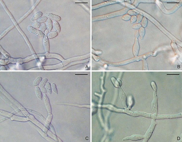
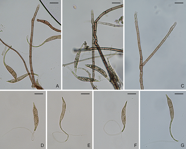
Figure. Nakataea serpens (NYBG985747). A–C. Conidiophores. D–G. Conidia. Scale bars: A–G = 20 µm.

Nakataea serpens Shearer & J.L. Crane, Trans. Br. mycol. Soc. 73(2): 370 (1979)
MycoBank: MB318483.

Hyphae branched, septate, light brown to brown, smooth, 1–3 µm diam. Conidiophores solitary, erect, straight or curved, unbranched, 4–9-septate, brown, smooth, 100–250 × 3.5–6.5 µm. Conidiogenous cells terminal, pale brown, 20–120 × 4–6 µm, with several protruding denticles, 1–2 × 1.5–2 µm. Conidia solitary, curved, smooth, 4-septate, not or slightly constricted at septum; main cells falcate, brown, 55–135 × 7–9 µm; apical cell sigmoid, hyaline to pale yellow, 80–140 × 1.5–3 µm. Sexual state unknown.

Typification: Holotype ILLS38410. Isotypes ILLS42909, NY985747.

Gene sequences: Unknown.

Specimens examined: USA, Illinois, Mason County, Quiver Creek, from plant debris, 11 Aug. 1978, C.A. Shearer, CS-612-1 (ILLS38410, ILLS42909, NY985747).

Hosts/substrates: On plant debris.

Distribution: USA (Illinois).

Copyright 2022 by The American Phytopathological Society. Reproduced, by permission, from Luo, J., and Zhang, N. 2022. The Rice Blast Fungus and Allied Species: A Monograph of the Fungal Order Magnaporthales (https://my.apsnet.org/APSStore/Product-Detail.aspx?WebsiteKey=2661527A-8D44-496C-A730-8CFEB6239BE7&iProductCode=46826). American Phytopathological Society, St. Paul, MN.

Magnaporthiopsis incrustans

Figure. Magnaporthiopsis incrustans (AF14SW17). A–D. Conidiophores and conidia. Scale bars: A–D = 10µm.

Magnaporthiopsis incrustans (Landsch. & N. Jacks.) J. Luo & N. Zhang, Mycologia 105(4): 1024 (2013).
MycoBank: MB802974.
        ≡ Gaeumannomyces incrustans Landsch. & N. Jacks., Mycol. Res. 93(1): 55 (1989).

Ascomata perithecial, immersed, solitary to gregarious, black, 179–420 µm diam, with a cylindrical, black neck, 252–672 × 84–147 µm. Paraphyses unbranched, septate, hyaline, dissolving at maturity. Asci 8-spored, unitunicate, clavate to cylindrical, 65–107 × 6–11 µm, with a refractive ring. Ascospores 3–4-seriate, fusiform, slightly curved, 3–5-septate, not constricted at septum, hyaline, smooth, 33–53 × 2–4 µm (Sexual state description from Landschoot and Jackson, 1989a). Asexual state phialophora-like. On CMA, hyphae branched, septate, hyaline, smooth, 2–5 µm diam. Conidiophores micronematous, solitary, erect, straight or curved, unbranched or sparsely branched, hyaline, smooth, 1–3-septate, 8–35 × 2–5 µm. Conidiogenous cells phialidic, erect, terminal or intercalary, hyaline, smooth, 8–17.5 × 3–6 µm. Conidia aggregated in slimy heads, ovoid to ellipsoidal, straight or slightly curved, aseptate, hyaline, smooth, 5–11 × 2–5 µm.

Colonies on PDA 6.5 cm diam after 7 days at 25 °C in dark; surface floccose, parrot green; aerial mycelium yellowish; reverse cedar green. Colonies on CMA reaching 6 cm after 7 days in dark at 25 °C; surface pale yellow-green; aerial mycelium sparse; reverse pale yellow-green.

Typification: Holotype DAR59042. Ex-holotype cultures ATCC64418, ATCC64416.

Gene sequences: JF414867 (18S), JF414843 (ITS), JF414892 (28S), JF710386 (MCM7), JF710437 (RPB1), JF710412 (TEF1).

Genome sequences: SRX795321 (genome), SRX798625 (transcriptome).

Specimens examined: USA, New Jersey, Adelphia Farm, on roots of Panicum virgatum, 8 Aug. 2014, J. Luo, RUTPPAF14SW17; ibid., New Brunswick, on roots of Festuca brevipila, 19 Jul. 2018, H. Chen and J. Murphy, RUTPPF2BHF1, RUTPPBHF9; Indiana, West Lafayette, on roots of Festuca, 15 Aug. 2014, T. Creswell, RUTPP1801540-11.

Hosts/substrates: From roots of Cynodon, Festuca, Poa annua, P. pratensis, and Zoysia (Poaceae).

Distribution: USA (Georgia, Indiana, Kansas, Maryland, Massachusetts, New Jersey, New York).

Copyright 2022 by The American Phytopathological Society. Reproduced, by permission, from Luo, J., and Zhang, N. 2022. The Rice Blast Fungus and Allied Species: A Monograph of the Fungal Order Magnaporthales (https://my.apsnet.org/APSStore/Product-Detail.aspx?WebsiteKey=2661527A-8D44-496C-A730-8CFEB6239BE7&iProductCode=46826). American Phytopathological Society, St. Paul, MN.

Gaeumannomyces mirabilis

Figure. Gaeumannomyces mirabilis (BPI880716). A. Ascomata. B–C. Asci. D–E. Ascospores. Scale bars: A = 500 µm; B–E = 10 µm.

Gaeumannomyces mirabilis (I. Hino & Katum.) Lar.N. Vassiljeva, Nizshie Rasteniya, Griby i Mokhoobraznye Dalnego Vostoka Rossii 4: 147. 1998.
MycoBank: MB 450454.
Diaboliumbilicus mirabilis I. Hino & Katum. [as ‘mirabile’], Bullettin of the Faculty of Agriculture, Yamaguchi University 6: 40. 1955.

Ascomata perithecial, immersed, solitary to gregarious, globose to subglobose, dark brown to black, 550–1000 µm diam, with a cylindrical, dark brown to black neck, 175–290 × 160–275 µm. Paraphyses septate, hyaline, dissolving at maturity. Asci 8-spored, unitunicate, clavate, 100–125 × 19–24.5 µm, with a refractive ring. Ascospores parallel in ascus, filiform to fusiform, not or slightly curved, 3–7-septate, not constricted at septum, hyaline to yellowish, smooth, 90–115 × 5.5–7.5 µm. Asexual state unknown.

Typification: Japan, on dead stems of Sasa veitchii (Poaceae).

Gene sequences: Unknown.

Specimens examined: Unknown, on dead stems of Bambusoideae, 5 Jan. 2009, I. Carden, BPI880716.

Hosts/substrates: On dead stems of Bambusoideae, such as Sasa veitchii (Poaceae).

Distribution: Japan.

References:
Hino I, Katumoto K. 1955. Illustrationes fungorum bambusicolorum. III. Bulletin of the Faculty of Agriculture Yamaguchi University. 6:29–68.
Vasilyeva LN. 1998. Nizshie Rasteniya, Griby i Mokhoobraznye Dalnego Vostoka Rossii. 4:1–419.

Geographical distribution: Japan.

Copyright 2022 by The American Phytopathological Society. Reproduced, by permission, from Luo, J., and Zhang, N. 2022. The Rice Blast Fungus and Allied Species: A Monograph of the Fungal Order Magnaporthales (https://my.apsnet.org/APSStore/Product-Detail.aspx?WebsiteKey=2661527A-8D44-496C-A730-8CFEB6239BE7&iProductCode=46826). American Phytopathological Society, St. Paul, MN.

Kohlmeyeriopsis medullaris

Figure. Kohlmeyeriopsis medullaris (NYBG1315597). A. Ascomatum. B–E. Asci. F–H. Ascospores. Scale bars: A = 100 µm; B = 40 µm; C–H = 20 µm.

Kohlmeyeriopsis medullaris (Kohlm., Volkm.-Kohlm. & O.E. Erikss.) Klaubauf, M.-H. Lebrun & Crous, Stud. Mycol. 79: 101 (2014).
MycoBank: MB810198.
        ≡ Gaeumannomyces medullaris Kohlm., Volkm.-Kohlm. & O.E. Erikss., Mycologia 87(4): 540 (1995).
= Trichocladium medullare (asexual state) Kohlm. & Volkm.-Kohlm., Mycotaxon 53: 349 (1995).

Ascomata perithecial, immersed, solitary, globose to subglobose, dark brown to black, 550–750 µm diam, with a dark brown to black, cylindrical neck, 150–250 × 120–150 µm. Paraphyses hyaline, septate. Asci 8-spored, unitunicate, cylindrical, 130–175 × 7–9 µm, with a refractive ring. Ascospores parallel in ascus, filiform, curved, 3–7-septate, not constricted at septum, hyaline to yellowish, smooth, 95–165 × 2.5–4 µm. Asexual state trichocladium-like. Hyphae branched, septate, hyaline to light brown, smooth. Conidiophores short, branched. Conidia two-celled, smooth; apical cell ellipsoidal, aseptate, dark brown, 13.5–20.5 × 7–10 µm; basal cell cylindrical or obconical, 0(–1) septate, light brown, 3.5–6 × 3–5 µm (Asexual state description from Kohlmeyer and Volkmann-Kohlmeyer, 1995).

Typification: Holotype IMSJK5528. Isotype NY1315597. Paratype, NY1315595. Ex-holotype culture CBS117849.

Specimens examined: USA, North Carolina, Carteret County, Broad Creek, on dead standing culms of Juncus roemerianus, 16 Mar. 1994, J. Kohlmeyer, 5528 (NY1315597, CBS117849); ibid., 28 Nov. 1993, J. Kohlmeyer, 5522 (NY1315595).

Gene sequences: KM484852 (ITS), KM484968 (28S), KM485068 (RPB1).

Hosts/substrates: On dead culms of Juncus roemerianus (Juncaceae).

Distribution: USA (North Carolina).

Copyright 2022 by The American Phytopathological Society. Reproduced, by permission, from Luo, J., and Zhang, N. 2022. The Rice Blast Fungus and Allied Species: A Monograph of the Fungal Order Magnaporthales (https://my.apsnet.org/APSStore/Product-Detail.aspx?WebsiteKey=2661527A-8D44-496C-A730-8CFEB6239BE7&iProductCode=46826). American Phytopathological Society, St. Paul, MN.

Ophioceras leptosporum

Figure. Ophioceras leptosporum (ILLS242). A. Ascomata. B–C. Asci. D–E. Ascospores. Scale bars: A = 200 µm; B–E = 10 µm.

Ophioceras leptosporum (S.H. Iqbal) J. Walker, Mycotaxon 11(1): 62 (1980).
MycoBank: MB113924.
        ≡ Gaeumannomyces leptosporus S.H. Iqbal, Trans. Br. mycol. Soc. 58(2): 346 (1972).

Ascomata perithecial, superficial to submerged, solitary to gregarious, globose, dark brown to black, 200–350 µm diam, with a filiform to cylindrical, dark brown to black neck, 800–2200 × 35–70 µm. Paraphyses unbranched, septate hyaline. Asci 8-spored, unitunicate, cylindrical, 75–95 × 5–7 µm, with a refractive ring. Ascospores parallel in ascus, filiform, 3–7-septate, not constricted at septum, hyaline to yellowish, smooth, 55–80 × 1.5–2 µm. Asexual state unknown.

Typification: Isotype ILLS242. Ex-isotype culture CBS894.70.

Gene sequences: JX134664 (18S), JX134678 (ITS), NG057959 (28S), JX134718 (MCM7), JX134732 (RPB1), JX134704 (TEF1).

Genome sequences: SRX798632 (transcriptome).

Specimens examined: UK, England, Exeter, River Creek near Cowley Bridge., on dead submerged stems of Apiaceae, 6 Feb. 1970, S.H. Iqbal, 57368 (ILLS242, culture CBS894.70). USA, Illinois, Union, Pine Hills Swamp Nature Preserve, on submerged, decayed stems, 5 May. 1977, J.L. Crane and J.D. Schoknecht 37175 (ILLS163261).

Hosts/substrates: On rotten, submerged stems and leaves.

Distribution: China, Malaysia, UK (England), USA (Illinois).

Copyright 2022 by The American Phytopathological Society. Reproduced, by permission, from Luo, J., and Zhang, N. 2022. The Rice Blast Fungus and Allied Species: A Monograph of the Fungal Order Magnaporthales (https://my.apsnet.org/APSStore/Product-Detail.aspx?WebsiteKey=2661527A-8D44-496C-A730-8CFEB6239BE7&iProductCode=46826). American Phytopathological Society, St. Paul, MN.

Ceratosphaeria pusilla


Figure. Ceratosphaeria pusilla (ILLS44364). A–B. Ascomata. C–D. Asci. E–G. Ascospores. Scale bars: A–B = 200 µm; C–G = 10 µm.

Ceratosphaeria pusilla (Fuckel) Sacc., Syll. fung. (Abellini) 2: 227 (1883).
MycoBank: MB241302.
        ≡ Ceratostoma pusillum Fuckel, Jb. nassau. Ver. Naturk. 23–24: 128 (1870).

Ascomata perithecial, superficial, solitary, ovoid to obpyriform, dark brown to black, 200–350 µm diam, with a black, conical to cylindrical neck, 330–450 × 60–90 µm. Paraphyses septate, hyaline. Asci 8-spored, unitunicate, cylindrical, 190–220 × 10–13 µm, with a refractive ring. Ascospores uniseriate in ascus, fusiform, 4–5-septate, slightly constricted at septum, hyaline, smooth, 25–30 × 6–9 µm. Asexual state unknown.

Typification: Unknown.

Specimens examined: USA, Illinois, Johnson, Elvira Cypress Swamp (Deer Pond), on rotten wood, 29 Nov. 1973, J.L. Crane and C.A. Shearer, 44364 (ILLS159659).

Gene sequences: Unknown.

Hosts/substrates: On rotten wood.

Distribution: Germany, Pakistan, USA (Illinois).

Copyright 2022 by The American Phytopathological Society. Reproduced, by permission, from Luo, J., and Zhang, N. 2022. The Rice Blast Fungus and Allied Species: A Monograph of the Fungal Order Magnaporthales (https://my.apsnet.org/APSStore/Product-Detail.aspx?WebsiteKey=2661527A-8D44-496C-A730-8CFEB6239BE7&iProductCode=46826). American Phytopathological Society, St. Paul, MN.

Ceratosphaeria mycophila

Figure. Ceratosphaeria mycophila (BPI624440). A. Ascomata. B–D. Asci. E–H. Ascospores. Scale bars: A = 200 µm; B–H = 10 µm.

Ceratosphaeria mycophila G. Winter, Rabenh. Krypt.-Fl., Edn 2 (Leipzig) 1.2: 258 (1885).
MycoBank: MB241375.

Ascomata perithecial, superficial, gregarious, globose, ovoid to obpyriform, dark brown to black, 350–850 µm diam, with a black, cylindrical neck. Paraphyses septate, hyaline. Asci 8-spored, unitunicate, cylindrical, 120–180 × 8–10 µm, with a refractive ring. Ascospores 2–4-seriate in ascus, filiform to fusiform, 5–7-septate, not constricted at septum, hyaline, smooth, 35–45 × 3–4.5 µm. Asexual state unknown.

Typification: Unknown.

Gene sequences: Unknown.

Specimens examined: USA, Wisconsin, Devil’s Lake, Devil’s Lake State Park, Sauk Co., on rotten wood, 4 Sep. 1953, W.W. Diehl, BPI624440.

Hosts/substrates: On rotten wood.

Distribution: UK, USA (Wisconsin).

Copyright 2022 by The American Phytopathological Society. Reproduced, by permission, from Luo, J., and Zhang, N. 2022. The Rice Blast Fungus and Allied Species: A Monograph of the Fungal Order Magnaporthales (https://my.apsnet.org/APSStore/Product-Detail.aspx?WebsiteKey=2661527A-8D44-496C-A730-8CFEB6239BE7&iProductCode=46826). American Phytopathological Society, St. Paul, MN.

Ceratosphaeria lampadophora

Figure. Ceratosphaeria lampadophora (ILLS159657). A–B. Ascomata. C–D. Asci. E–F. Ascospores. Scale bars: A–B = 200 µm; C–F = 10 µm.

Ceratosphaeria lampadophora (Berk. & Broome) Niessl, Verhandlungen des Naturforschenden Vereines in Brünn 14: 203 (1876).
MycoBank: MB250101.
        ≡ Sphaeria lampadophora Berk. & Broome, Annals and Magazine of Natural History 3: 372, t. 11: 28 (1859).

Ascomata perithecial, superficial, gregarious to solitary, dark brown to black, globose, ovoid to obpyriform, 350–850 µm diam, with a black, cylindrical neck. Paraphyses unbranched, septate, hyaline. Asci 8-spored, unitunicate, cylindrical, 135–175 × 8–10 µm, with a refractive ring. Ascospores 3–4-seriate in ascus, filiform to fusiform, 5–7-septate, not constricted at septum, hyaline, smooth, 60–75 × 3.5–4.5 µm. Asexual state unknown.

Typification: Broome, Oct. 1855; Broome, Apr. 1855; Pl. XI, fig. 28 (Annals and Magazine of Natural History 3).

Gene sequences: MN313785 (ITS), MN317269 (28S), MN313860 (ACT).

Specimens examined: USA, New Hampshire, Coos, White Mountain National Forest, Jefferson Notch Road at Jefferson Notch, on Betula, 1 Aug. 1983, J.L. Crane and J.D. Schoknecht, 58762 (ILLS159657).

Hosts/substrates: On wood.

Distribution: Australia, Canada (Ontario), Costa Rica (Alajuela, Cartago, Guanacaste, Limón, Puntarenas), Denmark (Sjælland), Ireland, Portugal (Braga), UK (Bath, Somersetshire). USA (New Hampshire, North Carolina, Tennessee, Wisconsin).

Copyright 2022 by The American Phytopathological Society. Reproduced, by permission, from Luo, J., and Zhang, N. 2022. The Rice Blast Fungus and Allied Species: A Monograph of the Fungal Order Magnaporthales (https://my.apsnet.org/APSStore/Product-Detail.aspx?WebsiteKey=2661527A-8D44-496C-A730-8CFEB6239BE7&iProductCode=46826). American Phytopathological Society, St. Paul, MN.

Ceratosphaeria grandis

Figure. Ceratosphaeria grandis (BPI625030). A. Ascomata. B–C. Asci. D–E. Ascospores. Scale bars: A = 500 µm; B–C = 20 µm; D–E = 10 µm.

Ceratosphaeria grandis Boud., Bulletin de la Société Mycologique de France 18: 143, tab. VII. fig. 2 (1902).
MycoBank: MB249855.

Ascomata perithecial, superficial, gregarious, ovoid to obpyriform, dark brown to black, 350–550 µm diam, covered with flexuous, thin-walled hairs. Paraphyses septate, hyaline. Asci 8-spored, unitunicate, clavate, 310–340 × 24–32 µm, with a refractive ring. Ascospores 2–4-seriate in ascus, cylindrical, sigmoid or geniculate, 5–6-septate, not constricted at septum, hyaline to yellowish, smooth, 55–75 × 5–7.5 µm. Asexual state unknown.

Typification: Unknown.

Gene sequences: Unknown.

Specimens examined: USA, California, Green Valley Falls, Solano County, on dead wood of Quercus agrifolia, 12 Feb. 1953, L. Bonar, BPI625030, BPI625031.

Hosts/substrates: On dead wood.

Distribution: France, USA (California).

Copyright 2022 by The American Phytopathological Society. Reproduced, by permission, from Luo, J., and Zhang, N. 2022. The Rice Blast Fungus and Allied Species: A Monograph of the Fungal Order Magnaporthales (https://my.apsnet.org/APSStore/Product-Detail.aspx?WebsiteKey=2661527A-8D44-496C-A730-8CFEB6239BE7&iProductCode=46826). American Phytopathological Society, St. Paul, MN.

Ceratosphaerella castillensis

Figure. Ceratosphaerella castillensis (NYBG968642). A. Ascomata. B–D. Asci. E–F. Ascospores. Scale bars: A = 500 µm; B–F = 10 µm.

Ceratosphaerella castillensis (C.L. Sm.) Huhndorf, Greif, Mugambi & A.N. Mill., Mycologia 100(6): 944 (2008).
MycoBank: MB508750.
        ≡ Ceratosphaeria castillensis C.L. Sm., Bull. Lab. Nat. Hist. Univ. Iowa 2:403. 1893.

Ascomata perithecial, superficial, gregarious, dark brown to black, 450–650 µm diam, with a cylindrical, dark brown to black neck, 500–1000 × 170–280 µm. Paraphyses unbranched, septate, hyaline. Asci 8-spored, unitunicate, clavate, 80–100 × 9–15 µm, with a refractive ring. Ascospores 2–4-seriate in ascus, fusiform, slightly curved, 3-septate, not constricted at septum, hyaline to yellowish, smooth, 29–35 × 3.5–5 µm.

Typification: Lectotype NY, C.L. Smith, Central American Fungi 13. Isotype BPI, C.L. Smith, Central American Fungi 13.

Gene sequences: EU528003 (18S), EU527997 (28S).

Specimens examined: Nicaragua, Castillo Viejo, on bark, Feb.–Mar. 1893, C.L. Smith, Central American Fungi 13 (NY968642, BPI625023, BPI625024).

Hosts/substrates: On bark or wood.

References:
Huhndorf SM, Greif M, Miller AN. 2008. Two new genera in the Magnaporthaceae, a new addition to Ceratosphaeria and two new species of Lentomitella. Mycologia. 100(6):940–955.

Geographical distribution: Costa Rica, Provincia Limon. Puerto Rico, Caribbean National Forest. Nicaragaua, Castillo Viejo.

Copyright 2022 by The American Phytopathological Society. Reproduced, by permission, from Luo, J., and Zhang, N. 2022. The Rice Blast Fungus and Allied Species: A Monograph of the Fungal Order Magnaporthales (https://my.apsnet.org/APSStore/Product-Detail.aspx?WebsiteKey=2661527A-8D44-496C-A730-8CFEB6239BE7&iProductCode=46826). American Phytopathological Society, St. Paul, MN.