Xenopyricularia zizaniicola

Xenopyricularia zizaniicola (Hashioka) Klaubauf, M.-H. Lebrun & Crous, Stud. Mycol. 79: 116 (2014).
MycoBank: MB810224.
        ≡ Pyricularia zizaniicola Hashioka [as ‘zizaniaecola‘], Trans. Mycol. Soc. Japan 14(3): 264 (1973).

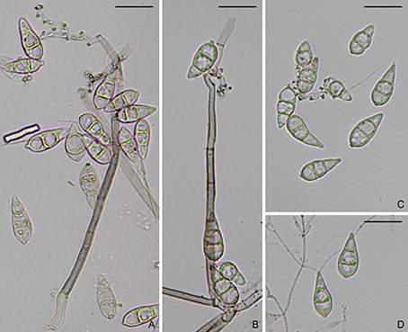
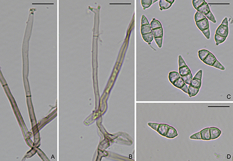
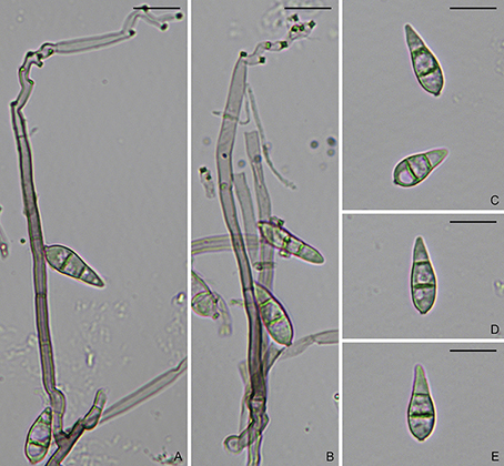
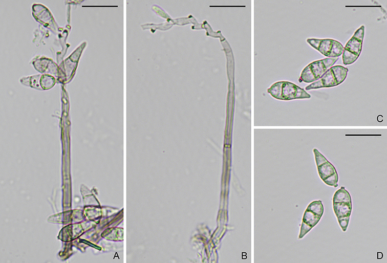
Asexual state pyricularia-like. On natural substrate, hyphae branched, septate, hyaline, smooth. Conidiophores solitary or gregarious, erect, curved, unbranched, subcylindrical, medium brown, 90–150(–220) × 4.5–5.2 µm. Conidiogenous cells integrated, terminal to intercalary, light brown, with several protruding denticles. Conidia solitary, ovate, 2-septate, light brown, 24–33 × 10.5–15.5 µm, with a truncate basal hilum. Appressoria subglobose, 12.1–10.5 µm. Sexual state unknown (Description from Hashioka, 1973).

Typification: Neotype CBSH-21846. Ex-neotype culture CBS133593.

Gene sequences: KM484947 (ITS), KM485042 (28S), KM485230 (ACT), AB274479 (CAL), KM485161 (RPB1), AB274463 (TUB).

Genome sequences: SRX798640 (transcriptome).

Hosts/substrates: On Zizania latifolia (Poaceae).

Distribution: Japan.

Copyright 2022 by The American Phytopathological Society. Reproduced, by permission, from Luo, J., and Zhang, N. 2022. The Rice Blast Fungus and Allied Species: A Monograph of the Fungal Order Magnaporthales (https://my.apsnet.org/APSStore/Product-Detail.aspx?WebsiteKey=2661527A-8D44-496C-A730-8CFEB6239BE7&iProductCode=46826). American Phytopathological Society, St. Paul, MN.

Pyricularia zingibericola

Figure. Pyricularia zingibericola (CBS138605). A–B. Conidiophores. C–D. Conidia. Scale bars: A–D = 20 µm.

Pyricularia zingibericola Klaubauf, M.-H. Lebrun & Crous, Stud. Mycol. 79: 115 (2014).
MycoBank: MB810222.

On CMA with sterile rice seeds, hyphae branched, septate, hyaline, smooth, 1.5–2 μm diam. Conidiophores solitary, erect, straight or curved, branched to unbranched, 3–7-septate, medium brown, smooth, 105–185 × 4–6 μm. Conidiogenous cells integrated, terminal or intercalary, light brown, smooth, 52–77 × 3–5 μm, with several protruding denticles, 1–1.5 × 1–2 µm. Conidia solitary, pyriform to obclavate, 2-septate, light brown, smooth, 18.5–26 × 8–10 μm, with a truncate hilum, 0.5–1.5 × 1–2 µm.

Colonies on 4.5 cm diam PDA after 7 days at 25 °C in dark; surface transparent with greenish olivaceous parts, white center, umbonate, powdery in center; reverse greenish olivaceous. Colonies on MEA 4 cm diam after 7 days at 25 °C in dark; surface transparent to white with leaden grey center, sulcate colony with entire edge, some irregular tufts; reverse pale with olivaceous grey center. Colonies on CMA 4 cm diam after 7 days at 25 °C in dark; surface grey olivaceous to olivaceous black with olivaceous grey center, entire edge, flat, slightly wool-like. Colonies on OA 4.5 cm diam after 7 days at 25 °C in dark; surface white with some dark spots, greenish olivaceous in center, flat, smooth, cotton-like (Description from Klaubauf et al., 2014).

Typification: Holotype CBSH-21845. Ex-holotype culture CBS138605 (RN0001).

Gene sequences: KM484941 (ITS), KM485037 (28S), KM485229 (ACT), KM485297 (CAL), KM485157 (RPB1).

Specimens examined: Réunion, on Zingiber officinale, J.C. Girard, culture CBS138605.

Hosts/substrates: On Zingiber officinale (Zingiberaceae).

Distribution: France (Réunion).

Copyright 2022 by The American Phytopathological Society. Reproduced, by permission, from Luo, J., and Zhang, N. 2022. The Rice Blast Fungus and Allied Species: A Monograph of the Fungal Order Magnaporthales (https://my.apsnet.org/APSStore/Product-Detail.aspx?WebsiteKey=2661527A-8D44-496C-A730-8CFEB6239BE7&iProductCode=46826). American Phytopathological Society, St. Paul, MN.

Pyricularia pennisetigena

Figure. Pyricularia pennisetigena (CBS138604). A–B. Conidiophores. C–D. Conidia. Scale bars: A–D = 20 µm.

Pyricularia pennisetigena Klaubauf, M.-H. Lebrun & Crous, Stud. Mycol. 79: 114 (2014).
MycoBank: MB810221.

On CMA with sterile rice seed, hyphae branched, septate, hyaline, smooth, 1.5–2.5 μm diam. Conidiophores solitary, erect, straight or curved, unbranched, 2–3-septate, medium brown, smooth, 85–150 × 3.5–6 μm. Conidiogenous cells integrated, terminal or intercalary, light brown, smooth, 30–88 × 3–5.5 μm, with several protruding denticles, 1–1.5 × 1–2 µm. Conidia solitary, pyriform to obclavate, 2-septate, light brown, smooth, 26–30.5 × 9–11 μm, with a truncate hilum, 0.5–1.5 × 1–2 µm.

Colonies on 5 cm diam PDA after 7 days at 25 °C in dark; surface fuscous black with grey center, broad white rim, flat, erose; reverse brown. Colonies on MEA 4.8 cm diam after 7 days at 25 °C in dark; surface cottony to velvety, buff, smoke grey, with broad white rim; reverse iron grey with pale margin. Colonies on CMA 5 cm diam after 7 days at 25 °C in dark; surface buff with grey dots. Colonies on OA 5 cm diam after 7 days at 25 °C in dark; surface buff (Description from Klaubauf et al., 2014).

Typification: Holotype CBSH21844. Ex-holotype culture CBS138604 (ML0036).

Gene sequences: KM484935 (ITS), KM485225 (ACT), KM485294 (CAL), KM485153 (RPB1).

Specimens examined: Mali, Cinzana, on Pennisetum, 19 Sep. 1990, J.L. Nottéghem, culture CBS138604.

Hosts/substrates: On Cenchrus ciliaris, C. echinatus, Echinochloa colona, and Pennisetum (Poaceae).

Distribution: Brazil, Japan, Mali, Philippines, USA.

Copyright 2022 by The American Phytopathological Society. Reproduced, by permission, from Luo, J., and Zhang, N. 2022. The Rice Blast Fungus and Allied Species: A Monograph of the Fungal Order Magnaporthales (https://my.apsnet.org/APSStore/Product-Detail.aspx?WebsiteKey=2661527A-8D44-496C-A730-8CFEB6239BE7&iProductCode=46826). American Phytopathological Society, St. Paul, MN.

Pyricularia penniseticola

Figure. Pyricularia penniseticola (CBS138603). A–B. Conidiophores. C–E. Conidia. Scale bars: A–E = 20 µm.

Pyricularia penniseticola Klaubauf, M.-H. Lebrun & Crous, Stud. Mycol. 79: 114 (2014).
MycoBank: MB810220.

On CMA with sterile rice seeds, hyphae branched, septate, hyaline, smooth, 1.5–2.5 μm diam. Conidiophores solitary, erect, straight or curved, branched, 2–5-septate, medium brown, smooth, 120–330 × 4–6 μm. Conidiogenous cells integrated, terminal or intercalary, light brown, smooth, 65–120 × 3–5.5 μm, with several protruding denticles, 1–1.5 × 1–2 µm. Conidia solitary, pyriform to obclavate, 2-septate, light brown, smooth, 25–33 × 9–10.5 μm, with a truncate hilum, 0.5–1.5 × 1–2 µm.

Colonies on 4.5 cm diam PDA after 7 days at 25 °C in dark; surface iron grey; reverse olivaceous black. Colonies on MEA 3 cm diam after 7 days at 25 °C in dark; surface light olivaceous grey; reverse olivaceous black. Colonies on CMA 3 cm diam after 7 days at 25 °C in dark. Colonies on OA 3.6 cm diam after 7 days at 25 °C in dark; surface sectoring (Description from Klaubauf et al., 2014).

Typification: Holotype CBSH21843. Ex-holotype culture CBS138603 (ML0031).

Gene sequences: KM484929 (ITS), KM485220 (ACT), KM485148 (RPB1).

Specimens examined: Mali, Segou field 2, from Digitaria exilis, 14 Sep. 1990, J.L. Nottéghem, culture CBS138603.

Hosts/substrates: On Digitaria exilis and Pennisetum (Poaceae).

Distribution: Burkina Faso, Ivory Coast, Mali.

Copyright 2022 by The American Phytopathological Society. Reproduced, by permission, from Luo, J., and Zhang, N. 2022. The Rice Blast Fungus and Allied Species: A Monograph of the Fungal Order Magnaporthales (https://my.apsnet.org/APSStore/Product-Detail.aspx?WebsiteKey=2661527A-8D44-496C-A730-8CFEB6239BE7&iProductCode=46826). American Phytopathological Society, St. Paul, MN.

Pyricularia ctenantheicola

Pyricularia ctenantheicola Klaubauf, M.-H. Lebrun & Crous, Stud. Mycol. 79: 111 (2014).
MycoBank: MB810219.

On CMA with sterile rice seeds, hyphae branched, septate, hyaline, smooth, 1.5–2 μm diam. Conidiophores solitary, erect, straight or curved, branched to unbranched, 1–6-septate, medium brown, smooth, 100–190 × 3–5.5 μm. Conidiogenous cells integrated, terminal or intercalary, light brown, smooth, 35–96 × 3–5 μm, with several protruding denticles, 1–2 × 1–2 µm. Conidia solitary, pyriform to obclavate, 2-septate, light brown, smooth to finely roughened, 20–25 × 7–9 μm, with a truncate hilum, 0.5–1.5 × 1–2 µm. Sexual state unknown.

Colonies on 3.5 cm diam PDA after 7 days at 25 °C in dark; surface hazel, with smoke grey tufts; reverse hazel. Colonies on MEA 2.7 cm diam after 7 days at 25 °C in dark; surface white to vinaceous buff, cottony, with undulating margin; reverse ochraceous to umber. Colonies on CMA 2.5 cm diam after 7 days at 25 °C in dark; surface pale luteous, with hazel center. Colonies on OA 3.5 cm diam after 7 days at 25 °C in dark (Description from Klaubauf et al., 2014).

Typification: Holotype CBSH21842. Ex-holotype culture CBS138601 (GR0002).

Gene sequences: KM484878 (ITS), KM484994 (28S), KM485182 (ACT), KM485252 (CAL), KM485098 (RPB1).

Specimens examined: Greece, Almyros, on Ctenanthe oppenheimiana, 1998, A.C. Pappas and E.J. Paplomatas, culture CBS138601.

Hosts/substrates: On Ctenanthe oppenheimiana (Marantaceae).

Distribution: Greece.

Copyright 2022 by The American Phytopathological Society. Reproduced, by permission, from Luo, J., and Zhang, N. 2022. The Rice Blast Fungus and Allied Species: A Monograph of the Fungal Order Magnaporthales (https://my.apsnet.org/APSStore/Product-Detail.aspx?WebsiteKey=2661527A-8D44-496C-A730-8CFEB6239BE7&iProductCode=46826). American Phytopathological Society, St. Paul, MN.

Magnaporthiopsis rhizophila

Magnaporthiopsis rhizophila (D.B. Scott & Deacon) J. Luo & N. Zhang, Mycologia 105(4): 1025 (2013).
MycoBank: MB802975.
        ≡ Magnaporthe rhizophila D.B. Scott & Deacon, Trans. Br. mycol. Soc. 81(1): 77 (1983).

Ascomata perithecial, superficial solitary to gregarious, black, 400–500 µm diam, with a cylindrical, black neck, up to 600 µm long, 100–200 µm wide. Paraphyses unbranched, septate, hyaline, dissolving at maturity. Asci 8-spored, unitunicate, clavate to cylindrical, 80–100 × 8–12 µm, with a refractive ring. Ascospores biseriate, fusiform, slightly curved, 3-septate, not constricted at septum, hyaline, smooth, 24–28 × 6–8 µm. Asexual state phialophora-like. On PDA, hyphae branched, septate, hyaline, brown to olivaceous brown, smooth, 3–5 µm diam. Conidiophores micronematous, solitary, erect, straight or curved, unbranched or sparsely branched, hyaline, smooth. Conidiogenous cells phialidic, erect, terminal or intercalary, hyaline, smooth, 6–18 × 3–6 µm. Conidia aggregated in slimy heads, ovoid to ellipsoidal, straight or slightly curved, aseptate, hyaline, smooth, 6–20 × 2–6 µm.

Colonies on PDA 8 cm diam after 10 days at 28 °C; surface thin, floccose, grey brown to olivaceous black; marginal hyphae curling back strongly towards center of colony (Description from Scott and Deacon, 1983).

Typification: Holotype PREM45952. Ex-holotype culture PREM45952.

Gene sequences: JF414858 (18S), JF414834 (ITS), JF414883 (28S), JF710384 (MCM7), JF710432 (RPB1), JF710408 (TEF1).

Genome sequences: SRX798599 (genome), SRX798627 (transcriptome).

Hosts/substrates: From roots of Cynodon dactylon, Panicum virgatum, Poa pratensis, and Triticum aestivum (Poaceae).

Distribution: Australia, South Africa, USA.

Notes: This species causes root disease of Cynodon dactylon (Bermuda grass), Poa pratensis (Kentucky bluegrass), and Triticum aestivum (wheat). It was also isolated from Panicum virgatum (switchgrass).

Copyright 2022 by The American Phytopathological Society. Reproduced, by permission, from Luo, J., and Zhang, N. 2022. The Rice Blast Fungus and Allied Species: A Monograph of the Fungal Order Magnaporthales (https://my.apsnet.org/APSStore/Product-Detail.aspx?WebsiteKey=2661527A-8D44-496C-A730-8CFEB6239BE7&iProductCode=46826). American Phytopathological Society, St. Paul, MN.

Magnaporthiopsis maydis

Magnaporthiopsis maydis (Samra, Sabet & Hing.) Klaubauf, M.-H. Lebrun & Crous, Stud. Mycol. 79: 101 (2014).
MycoBank: MB810225.
        ≡ Cephalosporium maydis Samra, Sabet & Hing., Phytopathology 53: 404 (1963).
        ≡ Harpophora maydis (Samra, Sabet & Hing.) W. Gams, Stud. Mycol. 45: 192 (2000).

Asexual state phialophora-like. On PDA, hyphae simple or branched, septate, hyaline, smooth. Conidiophores micronematous, solitary, erect, straight or curved, branched, hyaline, smooth, septate, 60–250 µm long. Conidiogenous cells phialidic, erect, terminal or intercalary, hyaline, smooth. Conidia aggregated in slimy heads, ovoid, ellipsoidal to oblong, straight or slightly curved, aseptate, hyaline, smooth, 3.6–14 × 3.3–3.6 µm. Sexual state unknown.

Colonies on MEA and PDA 9 cm diam after 9 days at 22–24 °C (Description from Samra et al., 1963).

Typification: From roots and stems of Zea mays and from soils, Egypt.

Gene sequences: KM009208 (18S), KM009160 (ITS), KM009148 (28S), KM009172 (MCM7), KM009184 (RPB1), KM009196 (TEF1).

Hosts/substrates: From roots and stems of Zea mays (Poaceae) and from soils.

Distribution: Egypt, Hungary, India, Israel, Portugal, Spain.

Notes: Samra et al. (1963) first described the fungus causing late wilt disease on maize in Egypt as Cephalosporium maydis. Based on ITS sequence and morphological characteristics, Gams (2000) transferred it to the asexual genus Harpophora in Magnaporthaceae. Klaubauf et al (2014) later reexamined the fungus and placed it in Magnaporthiopsis.

Copyright 2022 by The American Phytopathological Society. Reproduced, by permission, from Luo, J., and Zhang, N. 2022. The Rice Blast Fungus and Allied Species: A Monograph of the Fungal Order Magnaporthales (https://my.apsnet.org/APSStore/Product-Detail.aspx?WebsiteKey=2661527A-8D44-496C-A730-8CFEB6239BE7&iProductCode=46826). American Phytopathological Society, St. Paul, MN.

Gaeumannomyces graminis

Gaeumannomyces graminis (Sacc.) Arx & D.L. Olivier, Trans. Br. mycol. Soc. 35(1): 32 (1952).
MycoBank: MB297567.
Rhaphidophora graminis Sacc., Fungi venet. nov. vel. Crit., Ser. 2: 307 (1875).

        ≡ Ophiobolus graminis (Sacc.) Sacc., Reliq. Libert 2: no. 134 (1881).
        ≡ Ophiochaeta graminis (Sacc.) Hara, Journal of Plant Protection, Tokyo 3: 342 (1916).
        ≡ Gaeumannomyces graminis var. graminis (Sacc.) Arx & D.L. Olivier, Trans. Br. Mycol. Soc. 35: 32 (1952).
= Sphaeria cariceti Berk. & Broome, Ann. Mag. nat. Hist., Ser. 3 7: 455 (1861).
        ≡ Ophiobolus cariceti (Berk. & Broome) Sacc., Syll. fung. (Abellini) 2: 349 (1883).
        ≡ Linocarpon cariceti (Berk. & Broome) Petr., Sydowia 6: 387 (1952).
        ≡ Gaeumannomyces cariceti (Berk. & Broome) Lar.N. Vassiljeva, Nizshie Rasteniya, Griby i Mokhoobraznye Dalnego Vostoka Rossii, Griby.                Tom 4. Pirenomitsety i Lokuloaskomitsety (Sankt-Peterburg) 4: 146 (1998).

Morphological description: On MEA, mycelium consisting of septate, branched, smooth, hyaline to pale brown, 1–4 μm diam hyphae. Conidiophores differentiated, branched often verticillate, hyaline, pale brown to brown. Conidiogenous cells phialidic, solitary or grouped, terminal, hyaline to pale brown, cylindrical to lageniform, straight or curved, 7–30 × 1.5–4 μm, with a cylindrical to conical, refractive, collarette up to 3.5 μm long, 1–1.7 μm wide. Conidia lunate, allantoid, hyaline, 4–10 × 1–2 μm. Hyphopodia not observed (Hernández-Restrepo et al. 2016).

Culture characteristics: After 7 d at 25 °C: On PDA reaching 60 mm diam, aerial mycelium scarce to moderate, cottony, olivaceous grey, buff or isabelline, submerged mycelium darker, margin diffuse to rhizoid; reverse centre olivaceous grey, colourless to the periphery. On MEA reaching 62 mm diam, aerial mycelium abundant to moderate, cottony, pale olivaceous grey, darker to the periphery, submerged mycelium dark, margin effuse, rhizoid; reverse fuscous dark, rhizoid to the periphery. On OA reaching 62 mm diam, flat to cottony, greenish grey to grey olivaceous in the centre, white to colourless to the periphery, aerial mycelium moderate to abundant, white, submerged mycelium dark in the centre, margin effuse; reverse pale mouse grey (Hernández-Restrepo et al. 2016).

Specimens examined: Italy, Selva, Treviso, on Cynodon or Agropyron, Oct. 1874, slides as DAR 21032. USA, Florida, isolated from Cynodon dactylon × C. transvaalensis, 1987, M. Elliott, CPC26020 = CBS141384; 1991, M. Elliott, CPC26027; CPC26029; CPC26033 = CBS141385; CPC26035 = CBS141386; 1992, M. Elliott, CPC26039; CPC26042; CPC26045 (Hernández-Restrepo et al. 2016).

Pathogenicity: from Cynodon sp. or Agropyron sp.

References:
Hernández-Restrepo M, Groenewald JZ, Elliott ML, Canning G, McMillan VE, Crous PW. 2016. Take-all or nothing. Studies in Mycology 83:19–48.

Geographical distribution: Italy. USA, Florida.

Copyright 2022 by The American Phytopathological Society. Reproduced, by permission, from Luo, J., and Zhang, N. 2022. The Rice Blast Fungus and Allied Species: A Monograph of the Fungal Order Magnaporthales (https://my.apsnet.org/APSStore/Product-Detail.aspx?WebsiteKey=2661527A-8D44-496C-A730-8CFEB6239BE7&iProductCode=46826). American Phytopathological Society, St. Paul, MN.

Gaeumannomyces graminicola

Gaeumannomyces graminicola Hern.-Restr. & Crous, Stud. Mycol. 83: 38 (2016).
MycoBank: MB816896.

Morphological description: On MEA, mycelium consisting of septate, branched, smooth, hyaline to brown, 1–4 μm diam hyphae. Conidiophores more or less differentiated, verticillate. Conidiogenous cells phialidic, hyaline to pale brown, solitary or grouped, terminal, sometimes intercalary, cylindrical, lageniform, 5–20 × 2–4.5 μm, collarette up to 3 μm long, 1–2.5 μm diam. Conidia lunate, slightly or strongly curved, hyaline, 5–11.5 × 1–2 μm. Hyphopodia lobed, brown, 16.5–24 × 15.5–23.5 μm (Hernández-Restrepo et al. 2016).

Culture characteristics: After 7 d at 25 °C: On PDA reaching 74 mm diam, flat, aerial mycelium scarce, cottony, white, submerged mycelium dark, in the centre hazel, grey, isabelline, olivaceous grey, buff to the periphery, margin effuse, rhizoid; reverse fuscous black, mouse grey or isabelline in the centre, no change to the periphery. On MEA reaching 76 mm diam, aerial mycelium moderate, cottony to funiculose, white, mouse grey to pale mouse grey, submerged mycelium dark (mouse grey), margin effuse, rhizoid; reverse centre fuscous, periphery amber white to white. On OA reaching 77 mm diam, flat, aerial mycelium scarce to moderate or abundant, cottony to funiculose, white, submerged mycelium dark, olivaceous grey, olivaceous black, dark mouse grey, margin effuse, rhizoid; reverse olivaceous, mouse grey, leaden grey, no change to the periphery (Hernández-Restrepo et al. 2016).

Specimens examined: Netherlands, near Barendrecht, isolated from Ctenanthe, stem base, J.W. Veenbaas-Rijks (holotype, CBSH-22580; culture ex-type, CBS352.93). USA, Florida, isolated from Stenotaphrum secundatum, 1988, M. Elliott, CPC26022; 1990, M. Elliott, CPC26025 = CBS141381; 1991, M. Elliott, CPC26036 = CBS141382; Georgia, isolated from Eremochloa ophiuroides, 1994, H. Wilkinson, CPC26056 = CBS141383 (Hernández-Restrepo et al. 2016).

Hosts/substrates: from Ctenanthe sp., Stenotaphrum secundatum (buffalo grass), and Eremochloa ophiuroides (centipedegrass).

References:
Hernández-Restrepo M, Groenewald JZ, Elliott ML, Canning G, McMillan VE, Crous PW. 2016. Take-all or nothing. Studies in Mycology 83:19–48.

Geographical distribution: Netherlands. USA, Florida, Georgia.

Copyright 2022 by The American Phytopathological Society. Reproduced, by permission, from Luo, J., and Zhang, N. 2022. The Rice Blast Fungus and Allied Species: A Monograph of the Fungal Order Magnaporthales (https://my.apsnet.org/APSStore/Product-Detail.aspx?WebsiteKey=2661527A-8D44-496C-A730-8CFEB6239BE7&iProductCode=46826). American Phytopathological Society, St. Paul, MN.

Gaeumannomyces glycinicola

Gaeumannomyces glycinicola Hern.-Restr., G. Canning & Crous, Stud. Mycol. 83: 35 (2016).
MycoBank: MB816907.

Morphological description: On MEA, mycelium consisting of septate, branched, smooth, straight or flexuous, hyaline to brown, 1.5–4 μm diam hyphae. Hyphopodia hyaline getting dark brown when mature, lobed, 22.5–43 × 15–34 μm diam. Conidiophores and conidia not observed (Hernández-Restrepo et al. 2016).

Culture characteristics: After 7 d at 25 °C: On PDA reaching 90 mm diam, aerial mycelium scarce, white, submerged mycelium rhizoid, pale cinnamon, margin rhizoid; reverse pale cinnamon. On MEA reaching 70 mm diam, cottony, aerial mycelium abundant, dense, white, submerged umber, margin effuse; reverse interweave, umber. On OA reaching 90 mm diam, cottony, moderate and colourless (Hernández-Restrepo et al. 2016).

Specimens examined: USA, Indiana, isolated from Glycine max, 1974, D. Huber (holotype, CBSH-22579; culture ex-type, CPC26057 = DAR 28746); isolated from Glycine max (pods of soybean), 1974, unknown collector, CPC26266 = CBS141380. (Hernández-Restrepo et al. 2016).

Hosts/substrates: from Glycine max (soybean).

References:
Hernández-Restrepo M, Groenewald JZ, Elliott ML, Canning G, McMillan VE, Crous PW. 2016. Take-all or nothing. Studies in Mycology 83:19–48.

Geographical distribution: USA, Indiana.

Copyright 2022 by The American Phytopathological Society. Reproduced, by permission, from Luo, J., and Zhang, N. 2022. The Rice Blast Fungus and Allied Species: A Monograph of the Fungal Order Magnaporthales (https://my.apsnet.org/APSStore/Product-Detail.aspx?WebsiteKey=2661527A-8D44-496C-A730-8CFEB6239BE7&iProductCode=46826). American Phytopathological Society, St. Paul, MN.