Ceratosphaeria pusilla


Figure. Ceratosphaeria pusilla (ILLS44364). A–B. Ascomata. C–D. Asci. E–G. Ascospores. Scale bars: A–B = 200 µm; C–G = 10 µm.

Ceratosphaeria pusilla (Fuckel) Sacc., Syll. fung. (Abellini) 2: 227 (1883).
MycoBank: MB241302.
        ≡ Ceratostoma pusillum Fuckel, Jb. nassau. Ver. Naturk. 23–24: 128 (1870).

Ascomata perithecial, superficial, solitary, ovoid to obpyriform, dark brown to black, 200–350 µm diam, with a black, conical to cylindrical neck, 330–450 × 60–90 µm. Paraphyses septate, hyaline. Asci 8-spored, unitunicate, cylindrical, 190–220 × 10–13 µm, with a refractive ring. Ascospores uniseriate in ascus, fusiform, 4–5-septate, slightly constricted at septum, hyaline, smooth, 25–30 × 6–9 µm. Asexual state unknown.

Typification: Unknown.

Specimens examined: USA, Illinois, Johnson, Elvira Cypress Swamp (Deer Pond), on rotten wood, 29 Nov. 1973, J.L. Crane and C.A. Shearer, 44364 (ILLS159659).

Gene sequences: Unknown.

Hosts/substrates: On rotten wood.

Distribution: Germany, Pakistan, USA (Illinois).

Copyright 2022 by The American Phytopathological Society. Reproduced, by permission, from Luo, J., and Zhang, N. 2022. The Rice Blast Fungus and Allied Species: A Monograph of the Fungal Order Magnaporthales (https://my.apsnet.org/APSStore/Product-Detail.aspx?WebsiteKey=2661527A-8D44-496C-A730-8CFEB6239BE7&iProductCode=46826). American Phytopathological Society, St. Paul, MN.

Ceratosphaeria mycophila

Figure. Ceratosphaeria mycophila (BPI624440). A. Ascomata. B–D. Asci. E–H. Ascospores. Scale bars: A = 200 µm; B–H = 10 µm.

Ceratosphaeria mycophila G. Winter, Rabenh. Krypt.-Fl., Edn 2 (Leipzig) 1.2: 258 (1885).
MycoBank: MB241375.

Ascomata perithecial, superficial, gregarious, globose, ovoid to obpyriform, dark brown to black, 350–850 µm diam, with a black, cylindrical neck. Paraphyses septate, hyaline. Asci 8-spored, unitunicate, cylindrical, 120–180 × 8–10 µm, with a refractive ring. Ascospores 2–4-seriate in ascus, filiform to fusiform, 5–7-septate, not constricted at septum, hyaline, smooth, 35–45 × 3–4.5 µm. Asexual state unknown.

Typification: Unknown.

Gene sequences: Unknown.

Specimens examined: USA, Wisconsin, Devil’s Lake, Devil’s Lake State Park, Sauk Co., on rotten wood, 4 Sep. 1953, W.W. Diehl, BPI624440.

Hosts/substrates: On rotten wood.

Distribution: UK, USA (Wisconsin).

Copyright 2022 by The American Phytopathological Society. Reproduced, by permission, from Luo, J., and Zhang, N. 2022. The Rice Blast Fungus and Allied Species: A Monograph of the Fungal Order Magnaporthales (https://my.apsnet.org/APSStore/Product-Detail.aspx?WebsiteKey=2661527A-8D44-496C-A730-8CFEB6239BE7&iProductCode=46826). American Phytopathological Society, St. Paul, MN.

Ceratosphaeria lampadophora

Figure. Ceratosphaeria lampadophora (ILLS159657). A–B. Ascomata. C–D. Asci. E–F. Ascospores. Scale bars: A–B = 200 µm; C–F = 10 µm.

Ceratosphaeria lampadophora (Berk. & Broome) Niessl, Verhandlungen des Naturforschenden Vereines in Brünn 14: 203 (1876).
MycoBank: MB250101.
        ≡ Sphaeria lampadophora Berk. & Broome, Annals and Magazine of Natural History 3: 372, t. 11: 28 (1859).

Ascomata perithecial, superficial, gregarious to solitary, dark brown to black, globose, ovoid to obpyriform, 350–850 µm diam, with a black, cylindrical neck. Paraphyses unbranched, septate, hyaline. Asci 8-spored, unitunicate, cylindrical, 135–175 × 8–10 µm, with a refractive ring. Ascospores 3–4-seriate in ascus, filiform to fusiform, 5–7-septate, not constricted at septum, hyaline, smooth, 60–75 × 3.5–4.5 µm. Asexual state unknown.

Typification: Broome, Oct. 1855; Broome, Apr. 1855; Pl. XI, fig. 28 (Annals and Magazine of Natural History 3).

Gene sequences: MN313785 (ITS), MN317269 (28S), MN313860 (ACT).

Specimens examined: USA, New Hampshire, Coos, White Mountain National Forest, Jefferson Notch Road at Jefferson Notch, on Betula, 1 Aug. 1983, J.L. Crane and J.D. Schoknecht, 58762 (ILLS159657).

Hosts/substrates: On wood.

Distribution: Australia, Canada (Ontario), Costa Rica (Alajuela, Cartago, Guanacaste, Limón, Puntarenas), Denmark (Sjælland), Ireland, Portugal (Braga), UK (Bath, Somersetshire). USA (New Hampshire, North Carolina, Tennessee, Wisconsin).

Copyright 2022 by The American Phytopathological Society. Reproduced, by permission, from Luo, J., and Zhang, N. 2022. The Rice Blast Fungus and Allied Species: A Monograph of the Fungal Order Magnaporthales (https://my.apsnet.org/APSStore/Product-Detail.aspx?WebsiteKey=2661527A-8D44-496C-A730-8CFEB6239BE7&iProductCode=46826). American Phytopathological Society, St. Paul, MN.

Ceratosphaeria grandis

Figure. Ceratosphaeria grandis (BPI625030). A. Ascomata. B–C. Asci. D–E. Ascospores. Scale bars: A = 500 µm; B–C = 20 µm; D–E = 10 µm.

Ceratosphaeria grandis Boud., Bulletin de la Société Mycologique de France 18: 143, tab. VII. fig. 2 (1902).
MycoBank: MB249855.

Ascomata perithecial, superficial, gregarious, ovoid to obpyriform, dark brown to black, 350–550 µm diam, covered with flexuous, thin-walled hairs. Paraphyses septate, hyaline. Asci 8-spored, unitunicate, clavate, 310–340 × 24–32 µm, with a refractive ring. Ascospores 2–4-seriate in ascus, cylindrical, sigmoid or geniculate, 5–6-septate, not constricted at septum, hyaline to yellowish, smooth, 55–75 × 5–7.5 µm. Asexual state unknown.

Typification: Unknown.

Gene sequences: Unknown.

Specimens examined: USA, California, Green Valley Falls, Solano County, on dead wood of Quercus agrifolia, 12 Feb. 1953, L. Bonar, BPI625030, BPI625031.

Hosts/substrates: On dead wood.

Distribution: France, USA (California).

Copyright 2022 by The American Phytopathological Society. Reproduced, by permission, from Luo, J., and Zhang, N. 2022. The Rice Blast Fungus and Allied Species: A Monograph of the Fungal Order Magnaporthales (https://my.apsnet.org/APSStore/Product-Detail.aspx?WebsiteKey=2661527A-8D44-496C-A730-8CFEB6239BE7&iProductCode=46826). American Phytopathological Society, St. Paul, MN.

Ceratosphaerella castillensis

Figure. Ceratosphaerella castillensis (NYBG968642). A. Ascomata. B–D. Asci. E–F. Ascospores. Scale bars: A = 500 µm; B–F = 10 µm.

Ceratosphaerella castillensis (C.L. Sm.) Huhndorf, Greif, Mugambi & A.N. Mill., Mycologia 100(6): 944 (2008).
MycoBank: MB508750.
        ≡ Ceratosphaeria castillensis C.L. Sm., Bull. Lab. Nat. Hist. Univ. Iowa 2:403. 1893.

Ascomata perithecial, superficial, gregarious, dark brown to black, 450–650 µm diam, with a cylindrical, dark brown to black neck, 500–1000 × 170–280 µm. Paraphyses unbranched, septate, hyaline. Asci 8-spored, unitunicate, clavate, 80–100 × 9–15 µm, with a refractive ring. Ascospores 2–4-seriate in ascus, fusiform, slightly curved, 3-septate, not constricted at septum, hyaline to yellowish, smooth, 29–35 × 3.5–5 µm.

Typification: Lectotype NY, C.L. Smith, Central American Fungi 13. Isotype BPI, C.L. Smith, Central American Fungi 13.

Gene sequences: EU528003 (18S), EU527997 (28S).

Specimens examined: Nicaragua, Castillo Viejo, on bark, Feb.–Mar. 1893, C.L. Smith, Central American Fungi 13 (NY968642, BPI625023, BPI625024).

Hosts/substrates: On bark or wood.

References:
Huhndorf SM, Greif M, Miller AN. 2008. Two new genera in the Magnaporthaceae, a new addition to Ceratosphaeria and two new species of Lentomitella. Mycologia. 100(6):940–955.

Geographical distribution: Costa Rica, Provincia Limon. Puerto Rico, Caribbean National Forest. Nicaragaua, Castillo Viejo.

Copyright 2022 by The American Phytopathological Society. Reproduced, by permission, from Luo, J., and Zhang, N. 2022. The Rice Blast Fungus and Allied Species: A Monograph of the Fungal Order Magnaporthales (https://my.apsnet.org/APSStore/Product-Detail.aspx?WebsiteKey=2661527A-8D44-496C-A730-8CFEB6239BE7&iProductCode=46826). American Phytopathological Society, St. Paul, MN.

Buergenerula spartinae

Figure. Buergenerula spartinae (NYBG986085). A–B. Ascomata. C–D. Asci. E–F. Ascospores. G. Hyphopodium. Scale bars: A = 200 µm; B = 50 µm; C–G = 10 µm.

Buergenerula spartinae Kohlm. & R.V. Gessner, Can. J. Bot. 54(15): 1764 (1976).
MycoBank: MB310003.

Ascomata perithecial, immersed, solitary or gregarious, dark brown to black, 200–450 µm diam, with a cylindrical, hyaline to brownish neck, 150–250 × 90–150 µm. Paraphyses hyaline, septate, dissolving at maturity. Asci 8-spored, unitunicate, clavate, 100–165 × 15–20 µm, with a refractive ring. Ascospores biseriate in ascus, clavate, rounded at apex, narrowed toward base, straight or slightly curved, 3-septate, not or slightly constricted at septum, hyaline to yellowish, smooth, 35–55 × 9–13 µm. On CMA with seawater, conidiophores branched, up to 20 µm long. Conidiogenous cells phialidic. Conidia curved, 5 × 2 µm (Asexual state description from Kohlmeyer and Gessner, 1976).

Typification: Holotype NY J. Kohlmeyer (JK) No. 3498. Isotype IMS JK3498. Paratypes NY and IMS JK3503.

Specimens examined: Argentina, Buenos Aires, on Spartina alterniflora, 17 Oct. 1973, JK3498 (NY986083 notes, NY986084 dried specimen, NY986085 slides, NY986086 slides), JK3503 (NY986087).

Gene sequences: DQ341492 (28S), JX134666 (ITS), JX134692 (TEF1).

Genome sequences: SRX798618 (transcriptome).

Hosts/substrates: On leaf sheaths and dead stalks of Spartina and Panicum virgatum (Poaceae).

Distribution: Argentina, Canada, China, UK, USA.

Notes: Buergenerula spartinae was reported mainly on old leaf sheaths and dead stalks of Spartina in salt marshes.

Copyright 2022 by The American Phytopathological Society. Reproduced, by permission, from Luo, J., and Zhang, N. 2022. The Rice Blast Fungus and Allied Species: A Monograph of the Fungal Order Magnaporthales (https://my.apsnet.org/APSStore/Product-Detail.aspx?WebsiteKey=2661527A-8D44-496C-A730-8CFEB6239BE7&iProductCode=46826). American Phytopathological Society, St. Paul, MN. 

Bifusisporella

Bifusisporella sorghi R.M.F. Silva, R.J.V. Oliveira, J.D.P. Bezerra, Souza-Motta & G.A. Silva, Mycol. Progress 18: 852. 2019. (Type species).

On MEA, hyphae hyaline when young, light brown with age, occasionally producing structures similar to trapping loops, 2–3 μm diam. Conidiophores reduced to conidiogenous cells. Conidiogenous cells solitary or aggregate, curved, elongated, cylindrical or clavate, slightly curved at apex, light brown, 9.5–14.5 × 3–4 μm. Conidia dimorphic. Macroconidia curved, falcate, aseptate, guttulate, tapering to pointed ends, hyaline, smooth, 23–30 × 3–4 μm. Microconidia falcate, straight to slightly curved, hyaline, smooth, aseptate, produced either from a conidiogenous cell or directly from hyphae, 10–11.5 × 1–2 μm. Chlamydospores rarely observed, globose to subglobose, intercalary, terminal, single, light brown. Hyphopodia elongated, multi-lobulate, brown, smooth, 18–20 × 7.5–10 μm. Sexual state unknown (Description from Silva et al., 2019).

Typification: Holotype URM91354. Ex-holotype culture URM7442.

Gene sequences: MK060153 (28S), MK060155 (ITS), MK060157 (TEF1).

Host/substrate: On leaves of Sorghum bicolor (Poaceae).

Distribution: Brazil (Pernambuco).

How To Cite

Luo J, Zhang N. 2022. An E-monograph of the Fungal Order Magnaporthales: Taxonomy, Molecular Phylogeny, and Biogeography. https://magnaporthales.sebs.rutgers.edu.

Magnaporthiopsis poae

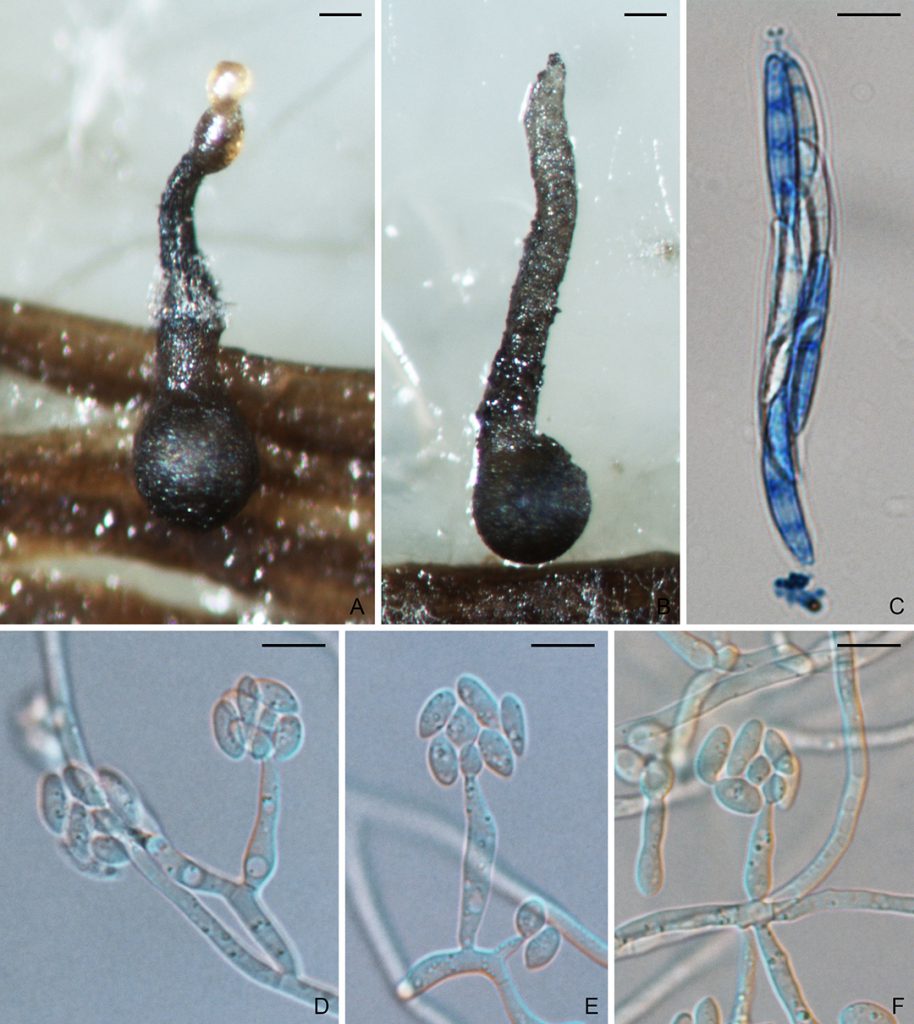
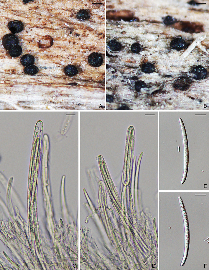
Figure. Magnaporthiopsis poae (M47 X M48). A–B. Ascomata. C. Ascus and ascospores. D–F. Conidiophores and conidia. Scale bars: A–B = 100um; C–F = 10µm.

Magnaporthiopsis poae (Landsch. & N. Jacks.) J. Luo & N. Zhang., Mycologia 105(4): 1024 (2013).
MycoBank: MB802973.
        ≡ Magnaporthe poae Landsch. & N. Jacks. Mycol. Res. 93:59 (1989).

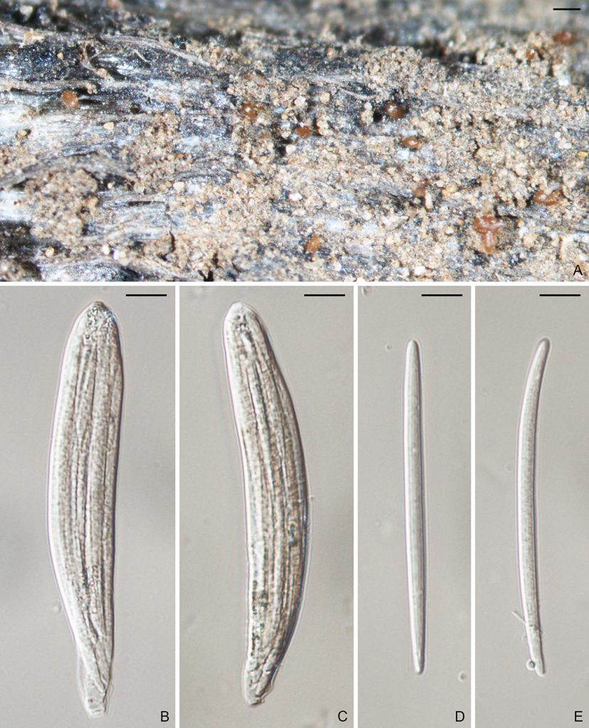
Ascomata perithecial, superficial or submerged, solitary or gregarious, dark brown to black, 230–500 µm diam, with a cylindrical, hyaline to brownish neck, 300–650 × 100–135 µm. Paraphyses septate, hyaline, dissolving at maturity. Asci 8-spored, unitunicate, clavate, 60–100 × 8–15 µm, with a refractive ring. Ascospores 1–4-seriate, fusiform, straight or slightly curved, 3-septate, not or slightly constricted at septum, unequally colored, smooth, 20–40 × 3.5–5.5 µm, with darkly pigmented inner cells. Asexual state phialophora-like. On CMA, hyphae branched, septate, hyaline to light brown, smooth, 2–5 µm diam. Conidiophores micronematous, solitary, erect, straight or curved, unbranched or sparsely branched, hyaline, smooth, 1–4-septate, 5–40 × 3–6.5 µm. Conidiogenous cells phialidic, erect, terminal or intercalary, hyaline, smooth, 5–20 × 2–6 µm. Conidia aggregated in slimy heads, ovoid to ellipsoidal, straight or slightly curved, aseptate, hyaline, smooth, 3–9 × 2–5 µm.

Colonies on PDA 7 cm diam after 7 days at 25 °C in dark; surface floccose, parrot green; aerial mycelium yellowish; reverse cedar green. Colonies on CMA reaching 6.5 cm after 7 days in dark at 25 °C; surface pale yellow green; aerial mycelium sparse; reverse pale yellow green.

Typification: Holotype DAR59044. Ex-holotype culture ATCC64411, ATCC64412.

Gene sequences: JF414860 (18S), JF414836 (ITS), JF414885 (28S), JF710390 (MCM7), JF710433 (RPB1), JF710415 (TEF1).

Genome sequences: ADBL01000000 (genome).

Specimens examined: USA, California, Nipomo, on Festuca, 19 Sep. 2018, D. Mosdell, RUTPP180919a3, RUTPP180919d1; New Jersey, New Brunswick, on Poa pratensis, 11 Aug. 2011, S. Zhao and L. Bierns, M47, M48; ibid., Adelphia, on Poa pratensis, 22 Aug. 2018, P.L. Vines, RUTPP11s-1, RUTPP1213-1, RUTPP14t-1, RUTPP1412-10, RUTPP1416-1; New York, on unknown host, 25 Aug. 2018, S. Tirpak and R. Buckley, RUTPPd29709-13.

Hosts/substrates: From Poaceae.

Distribution: Canada (Ontario), China, USA (California, New Jersey, New York).

Notes: Magnaporthiopsis poae infects turf roots, crowns, and leaf sheaths, and causes summer patch disease on Kentucky bluegrass (Poa pratensis), annual bluegrass (Poa annua), fine fescue (Festuca), perennial ryegrass (Lolium perenne), and bentgrass (Agrostis). The summer patch disease has been primarily reported in temperate regions of North America, ranging from Canada (Ontario) to USA from New Jersey and North Carolina as far west as California.

Copyright 2022 by The American Phytopathological Society. Reproduced, by permission, from Luo, J., and Zhang, N. 2022. The Rice Blast Fungus and Allied Species: A Monograph of the Fungal Order Magnaporthales (https://my.apsnet.org/APSStore/Product-Detail.aspx?WebsiteKey=2661527A-8D44-496C-A730-8CFEB6239BE7&iProductCode=46826). American Phytopathological Society, St. Paul, MN.

Gaeumannomyces tritici

Figure. Gaeumannomyces tritici (BPI 626756). A. Ascomata. B–C. Asci. D–E. Ascospores. Scale bars: A = 200um; B–E = 10µm.

Gaeumannomyces tritici (J. Walker) Hern.-Restr. & Crous, in Hernández-Restrepo, Groenewald, Elliott, Canning, Mcmillan & Crous, Stud. Mycol. 83: 26 (2016).
MycoBank: MB816900.
    ≡ Gaeumannomyces graminis var. tritici J. Walker, Trans. Br. mycol. Soc. 58(3): 439 (1972).

Morphological description: Ascomata perithecial, superficial or submerged, solitary or gregarious, dark brown to black, 200–450µm in diam, with a cylindrical, hyaline to brownish neck, 150–300 × 55–80 µm. Paraphyses hyaline, septate, dissolving at mature. Asci unitunicate, clavate, 8-spored, 90–120 × 10–18 µm, with a refractive ring. Ascospores parallel in the ascus, hyaline to yellowish, long fusiform to filiform, straight or slightly curved, 0–5-septate, not constricted at the septum, smooth, 70–110 × 3–5 µm.
    Conidiophores solitary, erect, straight or curved, unbranched or sparsely branched, hyaline, smooth. Conidiogenous cells phialidic, erect, terminal or intercalary, hyaline, smooth, 9–18 × 2–4 µm, with a cylindrical collarette, 1–3 × 1–2 µm. Conidia, lunate, allantoid to fusiform, straight to curved, hyaline, smooth, aseptate, 4–7 × 1–2 µm.

Specimens examined: Australia, New South Wales, on Triticum aestivum, 25 Feb. 1970, A.M. Smith, dar 19601 (BPI 626755); ibid., 12 Nov. 1969, L. Meakins, dar 19118 (BPI 626756). USA, New York, Geneva, on Triticum sativum, 5 Jul. 1921, R.J. Haskell and R.S. Kirby, BPI 626757.

Pathogenicity: Gaeumannomyces tritici can cause take-all diseases of wheat (Triticum), Barley (Hordeum), couch grass (Elymus repens), and wheatgrass (Agropyron) worldwide.

References:
Hernández-Restrepo M, Groenewald JZ, Elliott ML, Canning G, McMillan VE, Crous PW. 2016. Take-all or nothing. Studies in Mycology 83:19–48.

Geographical distribution: Australia, New South Walles. USA, New York.

Copyright 2022 by The American Phytopathological Society. Reproduced, by permission, from Luo, J., and Zhang, N. 2022. The Rice Blast Fungus and Allied Species: A Monograph of the Fungal Order Magnaporthales (https://my.apsnet.org/APSStore/Product-Detail.aspx?WebsiteKey=2661527A-8D44-496C-A730-8CFEB6239BE7&iProductCode=46826). American Phytopathological Society, St. Paul, MN.